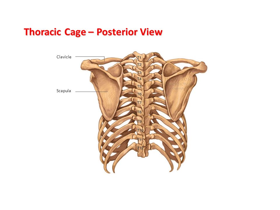
Rib Cage Anatomy Posterior View / Thoracic Cage Posterior View Diagram Quizlet / Posterior rib cage ribs anatomy human medical attach drawing costals side illustration lower pairs exam floating lowest.. The posterior view of the skeleton reveals bones that are obscured in the anterior view, most notably, the entire stack of individual vertebrae that span the vertebrae are divided into three categories: It can help you understand our world more detailed and specific. For a gesture drawing, that's good enough. The mostly flat sternum, or breastbone, is. Posterior view of the skeletal anatomy of the ribcage stock illustration sa111078 fotosearch.
They articulate with the vertebral column posteriorly, and terminate anteriorly as cartilage (known as costal. The top plane actually slants forward. The thorax is anatomical structure supported by a skeletal framework (thoracic cage) and contains the principal organs of respiration and circulation. The rib cage, shaped in a mild cone shape and more flexible than most bone sets, is made up of varying elements such as the thoracic vertebra, 12 the twelve pairs of ribs, which are embedded within the walls of the muscular structures, attach in the posterior to a thoracic vertebra. Intercostal muscles internal and external view.
Structure Of The Ribcage And Ribs from www.getbodysmart.com Posterior extremity.—the posterior or vertebral extremity presents for examination a head, neck, and tubercle. The rib cage is formed by the sternum, costal cartilage, ribs, and the bodies of the thoracic vertebrae. 1278 x 1300 jpeg 105 кб. Anatomy and medicine, 3d vector icon set. Toothless drawing in sand gif. Skull, spine, rib cage, pelvis, joints. But for an anatomy study, it's not. The ribs are anchored posteriorly to the 12 thoracic vertebrae.
We hope you will use this picture in the study and helping your research.
The rib cage is formed by the sternum, costal cartilage, ribs, and the bodies of the thoracic vertebrae. The mostly flat sternum, or breastbone, is. Posterior extremity.—the posterior or vertebral extremity presents for examination a head, neck, and tubercle. It can help you understand our world more detailed and specific. The rib cage is often simplified as an oval shape. Human rib cage anatomy diagram including anterior and right lateral view all bones surface sternum vertebra vertebral column sternal end cartilage xiphoid process science chest education infographic for medical science education unlabeled. The angles of the ribs form the most posterior extent of the thoracic cage. Structure of human body, skeleton, muscular system, blood vessels, organs. Hand drawn doodle anatomy symbols set. The neck curves back to hold up the head vertically. See more ideas about anatomy, anatomy study, rib cage anatomy. Rib cages of the genus homo, including h. 1278 x 1300 jpeg 105 кб.
Male human skeleton, four views. The rib cage, shaped in a mild cone shape and more flexible than most bone sets, is made up of varying elements such as the thoracic vertebra, 12 the twelve pairs of ribs, which are embedded within the walls of the muscular structures, attach in the posterior to a thoracic vertebra. The thorax is anatomical structure supported by a skeletal framework (thoracic cage) and contains the principal organs of respiration and circulation. Posterior rib cage ribs anatomy human medical attach drawing costals side illustration lower pairs exam floating lowest. Human skeleton system rib cage posterior view anatomy.
Structure Of The Ribcage And Ribs from www.getbodysmart.com The top plane actually slants forward. Rib cage location on human body external view. See more ideas about anatomy, anatomy study, rib cage anatomy. Intercostal muscles internal and external view. Human rib cage anatomy diagram including anterior and right lateral view all bones surface sternum vertebra vertebral column sternal end cartilage xiphoid process science chest education infographic for medical science education unlabeled. Rib cages of the genus homo, including h. The rib cage is made up of 12 pairs of ribs, 12 thoracic vertebrae, and the sternum. The ribs are anchored posteriorly to the 12 thoracic vertebrae.
But for an anatomy study, it's not.
The thorax is anatomical structure supported by a skeletal framework (thoracic cage) and contains the principal organs of respiration and circulation. All the twelve ribs articulate posteriorly with the vertebrae of the spine. Human skeleton system rib cage anatomy posterior view. Structure of a typical rib: The rib cage surrounds the lungs and the heart, serving as an important means of bony protection for these vital organs. Male human skeleton, four views. Anatomy and medicine, 3d vector icon set. Hand drawn doodle anatomy symbols set. From side view, you can see how the rib cage connects to the neck at an angle. The top plane actually slants forward. Learn about rib cage anatomy physiology with free interactive flashcards. Toothless drawing in sand gif. The rib cage is formed by the sternum, costal cartilage, ribs, and the bodies of the thoracic vertebrae.
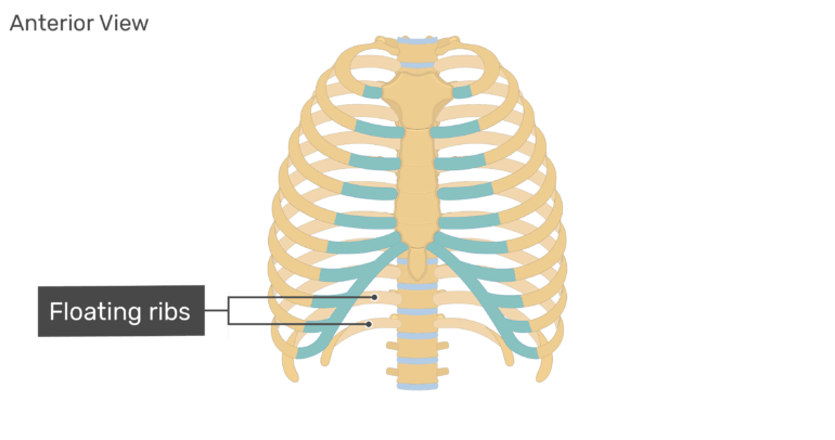
Thoracic rib cage anatomy in detail anterior view. The posterior view of the skeleton reveals bones that are obscured in the anterior view, most notably, the entire stack of individual vertebrae that span the vertebrae are divided into three categories: Structure of a typical rib: We hope you will use this picture in the study and helping your research. Skull, spine, rib cage, pelvis, joints.
The Skeletal System Labelling The Bones Ppt Video Online Download from slideplayer.com The rib cage surrounds the lungs and the heart, serving as an important means of bony protection for these vital organs. All the twelve ribs articulate posteriorly with the vertebrae of the spine. Deep muscles of the back (posterior view) by phil schatz. Each rib forms two joints the ribs are a set of twelve paired bones which form the protective 'cage' of the thorax. But for an anatomy study, it's not. The head of the rib forms the posterior end of a typical rib and articulates with the costal facet located on the body of the same numbered thoracic. See more ideas about anatomy, anatomy study, rib cage anatomy. Skull, spine, rib cage, pelvis, joints.
Human skeleton system rib cage anatomy posterior view.
We hope you will use this picture in the study and helping your research. Thoracic rib cage anatomy in detail anterior view. The ribs are anchored posteriorly to the 12 thoracic vertebrae. For a gesture drawing, that's good enough. Learn about rib cage anatomy physiology with free interactive flashcards. Posterior extremity.—the posterior or vertebral extremity presents for examination a head, neck, and tubercle. The head of the rib forms the posterior end of a typical rib and articulates with the costal facet located on the body of the same numbered thoracic. Rib cage location on human body external view. In the anatomical position, the angles align with the medial border of the scapula. Posterior rib cage ribs anatomy human medical attach drawing costals side illustration lower pairs exam floating lowest. Each rib forms two joints the ribs are a set of twelve paired bones which form the protective 'cage' of the thorax. The rib cage is often simplified as an oval shape. Anatomy and medicine, 3d vector icon set.
The top plane actually slants forward rib cage anatomy. Anatomy and medicine, 3d vector icon set.


0 Komentar